Low Free T3 With Normal TSH: When Thyroid Labs Miss the Energy Problem
Not all fatigue feels dramatic.
Sometimes it feels subtle.
You’re functioning.
You’re working.
You’re getting through the day.
But everything feels heavier than it should.
By early afternoon your clarity drops.
Your body feels colder than others around you.
Recovery from exercise takes longer.
Your motivation feels muted.
Your labs come back.
“Thyroid looks normal.”
But what was actually tested?
Usually just TSH.
And TSH does not produce energy.
Free T3 does.
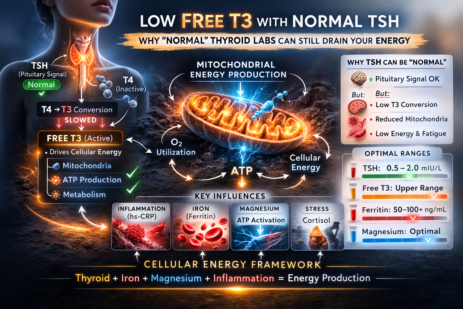
What TSH Actually Measures
TSH (thyroid-stimulating hormone) is produced by the pituitary gland.
It signals the thyroid to release hormone.
It does not measure:
-
Cellular thyroid activity
-
Mitochondrial density
-
ATP production
-
Peripheral hormone conversion
TSH reflects brain-to-thyroid communication.
It does not confirm what is happening inside your cells.
T4 vs T3: Only One Drives Cellular Energy
The thyroid primarily produces T4 (thyroxine).
T4 is largely inactive.
It must be converted into T3 by deiodinase enzymes in peripheral tissues.
Free T3 is the biologically active hormone that:
-
Enters cells
-
Binds nuclear receptors
-
Activates mitochondrial genes
-
Increases ATP production
If T4 → T3 conversion slows, Free T3 can fall into the lower portion of its reference range while TSH remains completely normal.
This creates the “normal labs, still exhausted” experience discussed in Why Your Blood Work Is Normal But You’re Exhausted.
Why Free T3 Matters for Mitochondrial Function
Free T3 regulates:
-
Electron transport chain protein expression
-
ATP synthase activity
-
Mitochondrial transcription factor A (TFAM)
-
PGC-1α (mitochondrial biogenesis regulator)
In simple terms:
Free T3 helps determine how many mitochondria you have — and how efficiently they function.
If Free T3 is suboptimal, mitochondrial output can decline.
For a deeper explanation of how mitochondria generate energy, review The Electron Transport Chain Explained Simply.
This is where thyroid signaling meets cellular ATP production.
Why Routine Thyroid Testing Misses This Pattern
Most screening panels include only TSH.
If it falls within the laboratory reference range, thyroid is labeled normal.
But reference ranges are designed to detect disease — not optimize performance.
Free T3 can sit in the lower third of its range and still be reported as normal.
For some individuals, that level may not support robust mitochondrial output.
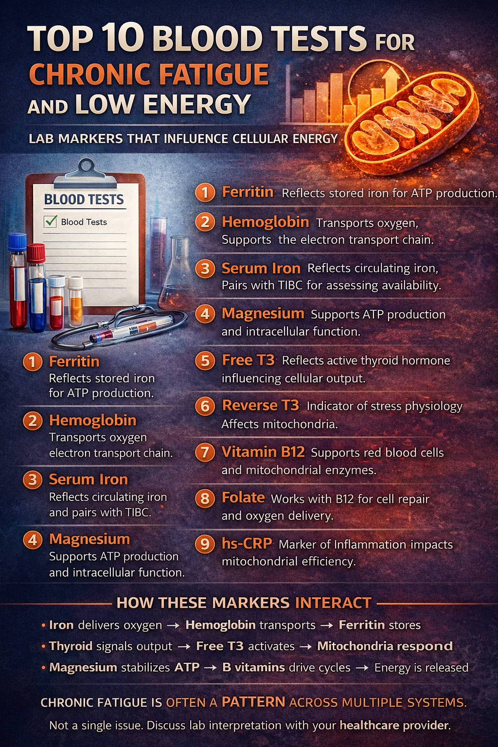
This is similar to the pattern described in Low Ferritin but Normal Hemoglobin — where oxygen transport appears adequate, but cellular reserves may be insufficient for optimal energy production.
What Lowers Free T3 Without Raising TSH?
Several stressors reduce peripheral conversion:
-
Chronic inflammation
-
Elevated hs-CRP
-
Caloric restriction
-
Chronic stress
-
Iron deficiency
-
Selenium deficiency
-
Insulin resistance
-
Recovery from illness
Inflammation is particularly important.
It suppresses deiodinase activity and increases reverse T3 production — an adaptive response that lowers metabolic rate.
When inflammation becomes chronic, conversion efficiency may remain suppressed.
The Thyroid–Iron–Magnesium Coordination Model
Thyroid does not operate alone.
Energy efficiency depends on coordination:
Iron supports electron transfer inside mitochondria.
Thyroid hormone increases mitochondrial protein expression.
Magnesium activates ATP so it can be used.
If Free T3 is low-normal and ferritin is also low, energy production may decline from two directions:
Reduced mitochondrial activation
Reduced iron-dependent electron transport
If magnesium is suboptimal, ATP activation may weaken further.
For context on ATP activation, review Magnesium and ATP: Why It Matters for Energy and Fatigue.
This is not a single-marker issue.
It is a systems pattern.
Where This Fits Inside the Larger Framework
Within the Cellular Energy Framework, thyroid signaling regulates the Thyroid–Mitochondria Axis.
Free T3 influences:
-
Basal metabolic rate
-
Oxygen utilization
-
Mitochondrial density
-
Electron transport chain efficiency
When multiple systems sit at mildly suboptimal levels simultaneously, fatigue often appears long before labs fall outside reference ranges.
For a structured overview of evaluating patterns instead of isolated numbers, see the Educational Blood Lab Interpretation Guide.
When to Discuss This With Your Healthcare Provider
If you have:
-
Normal TSH
-
Normal Free T4
-
Low-normal Free T3
-
Persistent fatigue
You may consider discussing:
-
Full thyroid panel
-
Ferritin and iron status
-
hs-CRP
-
Magnesium status
-
Selenium intake
This article is educational and not a substitute for medical care.
Thyroid treatment decisions should always be made with a licensed healthcare professional.
Frequently Asked Questions
Can you have thyroid symptoms with normal TSH?
Yes. TSH reflects pituitary signaling, not tissue-level thyroid activity. Free T3 drives mitochondrial energy production inside cells.
What is the difference between T4 and T3?
T4 is largely inactive. It must be converted into T3, the active hormone that regulates mitochondrial function and metabolism.
Can low Free T3 cause fatigue?
Yes. Free T3 regulates mitochondrial biogenesis and ATP production. Suboptimal levels may reduce cellular energy output even if TSH is normal.
How does inflammation affect thyroid conversion?
Inflammation suppresses deiodinase enzymes that convert T4 into T3. It may increase reverse T3, reducing active thyroid signaling.
Should everyone test Free T3?
Not necessarily. Pattern recognition matters more than ordering every possible marker. A thyroid panel, iron studies, magnesium, and hs-CRP often provide sufficient context when interpreted together.
