7 Blood Markers That Signal Mitochondrial Stress in Lee’s Summit Residents
Many residents in Lee’s Summit and the greater Kansas City metropolitan area search for answers when persistent fatigue, brain fog, low motivation, or poor recovery continue—even though routine blood work appears “normal.”
This situation can be frustrating. Standard laboratory testing is designed primarily to detect overt disease, but subtle metabolic stress often appears earlier through patterns in everyday laboratory markers.
One of the most important systems involved in energy production is the mitochondria.
These tiny structures inside cells function as the body’s cellular power plants, producing ATP—the molecule that fuels nearly every biological process in the body.
When mitochondria experience stress due to nutrient imbalances, inflammation, metabolic strain, or chronic stress, energy efficiency can decline long before major laboratory abnormalities appear.
Understanding these early signals may help explain why symptoms such as fatigue or brain fog sometimes appear even while blood tests remain within reference ranges.
For a broader overview of these concepts, see Functional Medicine in Lee’s Summit.
Why Mitochondrial Stress Matters in Lee’s Summit
Daily life in Lee’s Summit, Overland Park, Blue Springs, and the Kansas City metro region places significant demands on the body’s energy systems.
Busy schedules, professional responsibilities, commuting, family obligations, and ongoing stress all increase the metabolic workload placed on mitochondria.
When mitochondrial efficiency declines, individuals may begin to notice symptoms such as:
-
persistent fatigue
-
brain fog
-
slower physical recovery
-
reduced motivation
-
decreased mental clarity
Routine laboratory testing often measures circulating chemistry, but it may not fully reflect what is occurring inside cells.
This is why examining patterns across multiple markers may sometimes provide insight into metabolic stress before obvious abnormalities develop.
To better understand how the body produces energy at the cellular level, explore the Cellular Energy Framework.
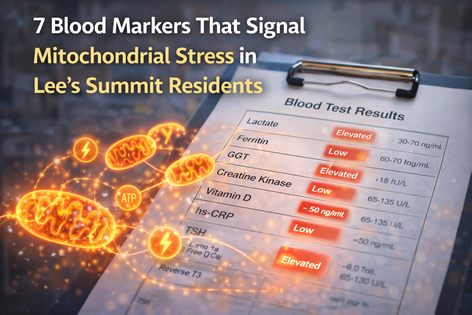
7 Blood Markers That Often Signal Mitochondrial Stress
The following laboratory markers sometimes reveal patterns associated with mitochondrial strain, particularly when interpreted together rather than individually.
These markers alone are not diagnostic but may provide useful context when viewed as part of a broader metabolic picture.
1. Elevated or High-Normal Lactate
Lactate levels may rise when mitochondria are unable to fully process pyruvate through the Krebs cycle and oxidative phosphorylation pathways.
When this occurs, cells shift toward less efficient anaerobic metabolism, which produces less ATP.
Even high-normal lactate levels may indicate metabolic stress in individuals experiencing fatigue or reduced exercise tolerance.
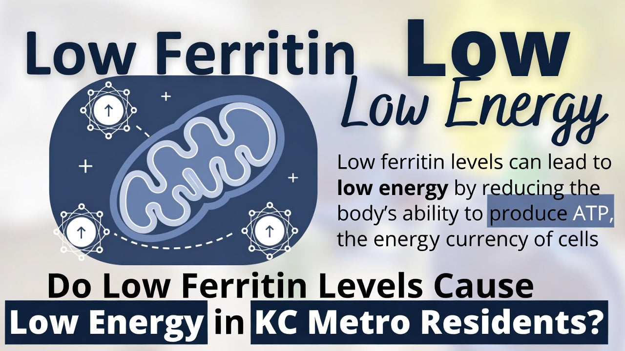
2. Low or Borderline Ferritin
Iron plays a key role in the electron transport chain, where mitochondria generate ATP.
Ferritin levels within the lower portion of the reference range may sometimes correlate with reduced cellular energy production, even when hemoglobin remains normal.
Lower iron reserves may limit oxygen utilization within mitochondria and contribute to fatigue.
For more information on how nutrients influence metabolism, see the Metabolic Nutrient Framework.
3. Elevated Gamma-Glutamyl Transferase (GGT)
GGT is commonly associated with liver function, but it can also reflect oxidative stress and glutathione demand.
Mitochondria rely heavily on antioxidant protection to maintain efficiency. When oxidative stress increases, mitochondrial workload rises and energy production may decline.
4. Abnormal Creatine Kinase (CK)
Creatine kinase reflects muscle energy metabolism and cellular turnover.
Two patterns sometimes observed include:
Low CK levels, suggesting reduced muscle energy turnover
High CK levels, suggesting increased cellular stress or muscle strain
Both patterns may appear in individuals experiencing persistent fatigue.
5. Low Vitamin D
Vitamin D influences several physiological systems involved in energy production, including:
-
mitochondrial respiration
-
immune regulation
-
inflammation balance
-
muscle function
Suboptimal vitamin D levels have been associated with fatigue and decreased physical performance.
6. Elevated Inflammatory Markers
Low-grade chronic inflammation places additional stress on mitochondrial systems.
Inflammatory signals increase oxidative stress and metabolic demand, potentially reducing mitochondrial efficiency and contributing to fatigue or brain fog.
7. Thyroid Conversion Patterns
Thyroid hormones regulate metabolic rate and mitochondrial activity.
Sometimes laboratory patterns show normal TSH levels while Free T3 remains low within the range or Reverse T3 appears elevated.
These patterns may reflect reduced thyroid hormone conversion, which can influence cellular energy production even when standard thyroid markers appear normal.
For more information on fatigue-related lab patterns, see Blood Test Markers That Affect Energy, Fatigue, and Brain Fog.
Why These Markers Often Remain “Normal”
Laboratory reference ranges are designed primarily to detect disease states, not necessarily optimal metabolic function.
The body has significant compensatory capacity, meaning symptoms may develop while laboratory values remain within standard ranges.
This gap between symptoms and laboratory interpretation is one reason many individuals in Lee’s Summit and the Kansas City metro region begin researching deeper explanations for fatigue and brain fog.
To understand how lab markers can be interpreted together, see Educational Blood Lab Interpretation and Optimal vs Standard Lab Ranges.
Functional Medicine and Mitochondrial Health in Lee’s Summit
Many individuals exploring persistent fatigue encounter the concept of functional medicine.
Functional medicine often examines health through a systems-biology perspective, looking at how nutrition, stress, sleep, metabolism, and inflammation interact.
Residents researching functional medicine in Lee’s Summit are often seeking a deeper explanation for why symptoms persist even when laboratory tests appear normal.
CelluShine focuses on educational approaches that help individuals understand how laboratory markers may relate to cellular metabolism and energy production pathways.
CelluShine provides educational tools to decode these patterns—not medical diagnosis or treatment.
For a complete overview of this educational framework, explore Functional Medicine in Lee’s Summit.
Looking Beyond Individual Lab Markers
No single laboratory marker explains fatigue.
Energy metabolism depends on multiple biological systems interacting together, which is why examining patterns across markers may provide greater insight than looking at individual values alone.
For additional information, see Mitochondrial Dysfunction and Hydration & Electrolytes.
Frequently Asked Questions
What blood markers can signal mitochondrial stress?
Markers such as lactate, ferritin, GGT, creatine kinase, vitamin D, inflammatory markers, and thyroid hormone patterns may sometimes reflect mitochondrial stress when viewed together.
Why do symptoms appear before laboratory abnormalities?
The body can compensate for metabolic stress for extended periods of time. Symptoms may develop while laboratory values remain within standard ranges.
How can mitochondrial stress contribute to brain fog?
The brain consumes a large portion of the body’s energy supply. Reduced ATP production can affect cognitive clarity and mental stamina.
Why are Lee’s Summit residents researching functional medicine?
Many individuals explore functional medicine concepts when persistent symptoms such as fatigue or brain fog are not fully explained by conventional testing.
References
Institute for Functional Medicine
National Institutes of Health – Mitochondrial Function
Wallace DC. Mitochondrial bioenergetics in health and disease
Filler K et al. Association of mitochondrial dysfunction and fatigue
Finsterer J. Biomarkers for detecting mitochondrial disorders
Hubens WHG et al. Blood biomarkers for mitochondrial dysfunction
Continue Reading in the CelluShine Education Series
Explore additional articles in this series:
Why Lee’s Summit Residents Feel Exhausted Despite “Normal” Blood Tests
Mitochondrial Dysfunction: The Hidden Cause of Fatigue in Lee’s Summit
